


As soon as you visit our department, you will be approached by a dedicated team of specialists who will tailor the exams to your individual needs. We take advantage of the all the benefits of modern imaging through advanced MRI scans. Our aim is state-of-the-art imaging, adjusted to the needs of each patient individually.
The latest MRI techniques are performed by doctors who have received training in centers of excellence in the USA. Image optimization is achieved in partnership with a qualified team of radiophysicists and biomedical engineers. The scientific team is further supported by fully qualified radiologic technologists, as well as nursing and administrative staff.
In line with international standards, our Unit works closely with the surgery clinics (Neurosurgery, Urology, Breast Center), as well as the Medical Oncology and Radiation Oncology Clinics. For improved individualized treatment of each patient and optimal diagnostic and treatment result, the imaging information offered by the scans performed within the Unit is presented at the weekly Metropolitan Hospital Oncology and Neurosurgery Councils.
What Is An MRI
Magnetic Resonance Imaging (MRI) is a non-invasive imaging method that combines the properties of magnetic fields and radiofrequencies to compose detailed images of the human body organs.
How Does It Work
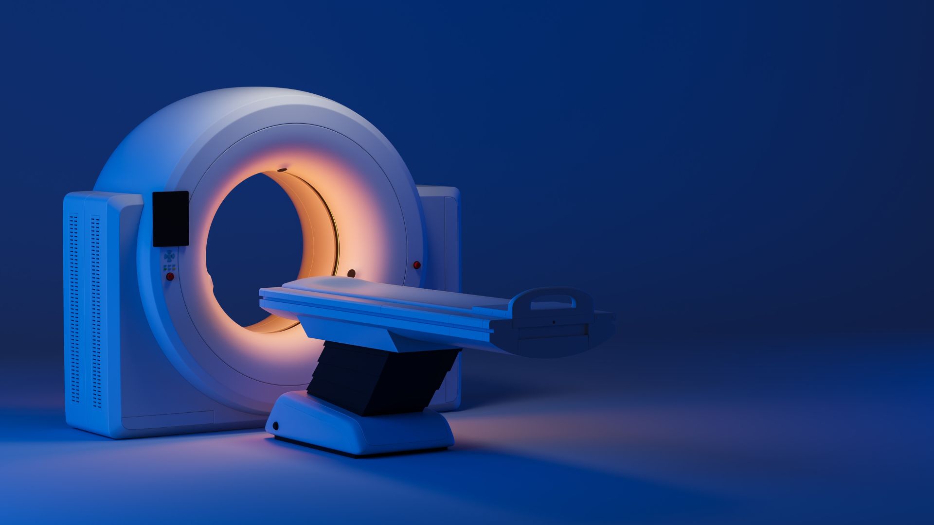
The MRI scanner is a large cylindrical machine, which is open on both sides. A magnetic field develops within the cylinder. When combined with radiofrequencies, the field temporarily shifts the hydrogen atoms within the human body. Upon returning to their original position, these atoms emit radiofrequencies. The signal is picked up by special computers, which process it and convert it into an image. Since the method does not use radiation, it is perfectly safe.
Do I Need To Prepare In Some Way?
Patients are placed inside the cylinder head or feet first, on their back or front (breast), depending on the anatomical area that needs to be examined.
No special preparation is required for central nervous system or breast scans. For pelvic scans, patients are advised to have a light meal 4 hours before the exam. Special instructions may be given in some cases, depending on the anatomical area and the clinical question.
Is The Contrast Medium Necessary
In many cases it is necessary to administer a contrast medium for MRIs (gadolinium), to get the necessary information from the organ being investigated. This contrast medium is different to the one used for CT scans. It does not contain iodine and serious allergic reactions are rare.